
digital xray
- Home
- Digital xray
Digital radiography is a type of X-ray imaging that uses digital X-ray sensors to replace traditional photographic X-ray film, producing enhanced computer images of teeth, gums, and other oral structures and conditions.
Digital dental images are acquired through three methods: the direct method, indirect method and semi-indirect method. The direct method uses an electronic sensor placed in the mouth to record images. The indirect technique uses an X-ray film scanner to view traditional dental X-rays as digital images. The semi-indirect digital technique combines a sensor and scanner to convert dental X-rays into digital film.
The Benefits and Advantages of Digital Dental X-Rays
The dental industry is ever-changing and adapting as new technology emerges to fit the needs of dentists and their patients. One way that dentistry has seen great advancements is through better and more advanced imaging processes.
Types and Uses of Digital Dental Radiographs
Digital dental radiographs can be taken inside (intraoral) or outside (extraoral) the mouth. Intraoral X-rays, the most commonly taken dental X-ray, provide great detail and are used to detect cavities, check the status of developing teeth and monitor teeth and bone health. Extraoral X-rays do not provide the detail of intraoral X-rays and are not used to identify individual tooth problems. However, they are used to detect impacted teeth, monitor jaw growth and development, and identify potential problems between teeth, jaws and temporomandibular joints (TMJ), or other facial bones. Types of intraoral X-rays include: 1.Bitewing X-rays, which are taken with the patient biting down on film, show details of the upper and lower teeth in one area of the mouth. Each bitewing shows a tooth from its crown (top) to about the level of the supporting bone. Bitewing X-rays are used to detect decay between teeth and changes in bone density caused by gum disease, as well as to determine the fit of dental crowns or restorations, and the marginal integrity of tooth fillings. 2.Periapical (limited) X-rays show the whole tooth from the crown to beyond the root tips to the supporting bone in one area of either the upper or lower jaw. Periapical X-rays are used to detect root structure and surrounding bone structure abnormalities. Showing bone loss around each tooth, periapical X-rays aid in treating conditions such as periodontitis, advanced gum disease, and detecting endodontic lesions (abscess).Types of extraoral X-rays include:
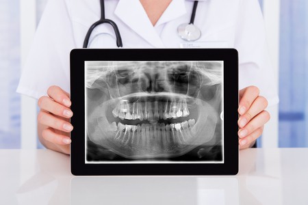
1.Panoramic (Panorex) X-rays
which require a machine that rotates around the head, show the entire mouth, including all the teeth in the upper and lower arch, in one image. Panoramic X-rays are used to plan treatment for dental implants, detect impacted wisdom teeth and jaw problems, and diagnose bony tumors and cysts. Panoramic films are used for forensic and legal purposes to identify otherwise unrecognizable bodies after fires, crashes or other fatalities.
2.Multi-slice computed tomography (MCT)
shows a particular layer or “slice” of the mouth while blurring all other layers. This type of X-ray is useful for examining structures that are difficult to see clearly.
3.Cephalometric projections
which show the entire head, help examine teeth in relation to a patient’s jaw and profile. Orthodontists, specialists in aligning and straightening teeth, use cephalometric projections to develop their treatment plans.
4.Sialography
uses a dye (radiopaque contrast agent) injected into the salivary glands to visualize them on the X-ray film. A sialography typically is used to identify salivary gland problems, such as blockages or Sjogren’s syndrome, an autoimmune disease that impedes saliva and tear production.
Cone beam computerized tomography (CBCT)
shows the body’s interior structures as a three-dimensional image. CBCT — often performed in a hospital or imaging center, but increasingly being used in the dental office — is used to identify facial bone problems, such as tumors or fractures. CT scans also are used to evaluate bone for dental implant placement and difficult tooth extractions to avoid possible complications during and after surgical procedures.